01 / CLINICAL HISTORY
More than 15 years of pain, tinnitus and restricted mandibular opening.
In 2002, a 46-year-old patient presented to the Oral and Maxillofacial Surgery service with temporomandibular joint pain of more than 15 years' duration. The surviving record also identifies tinnitus and limitation of mouth opening.
During that prolonged course, the patient had undergone occlusal rehabilitation, acrylic splint therapy and treatment recorded as “Miotens,” without satisfactory clinical improvement. The archive does not preserve standardized pain scores, measured maximal interincisal opening, magnetic-resonance imaging, or the precise sequence and duration of those earlier therapies.
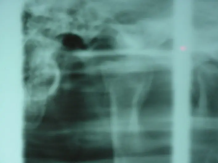
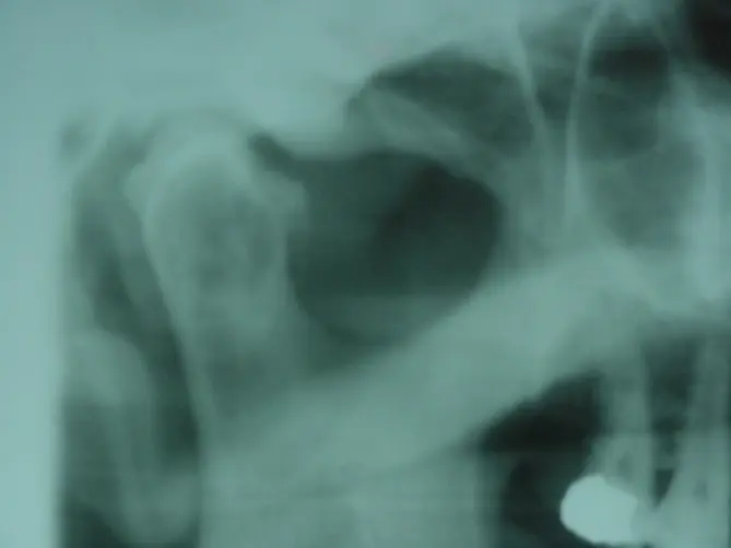
The treating team considered the bilateral osseous deformity mechanically relevant to joint function. Modern terminology would require clinical and cross-sectional imaging correlation before assigning a specific degenerative diagnosis; the historical record is therefore presented without retrospectively converting the finding into a diagnosis not explicitly documented.
02 / OPERATIVE DECISION
The goal was to restore a smoother condylar contour while preserving the joint.
After occlusal rehabilitation and stabilization, the team planned bilateral condylar arthroplasty through preauricular approaches. The operative concept was conservative in anatomical scope: expose both joints, identify the deforming exostotic projections, and selectively recontour the condyles rather than replace the joints.
This choice followed a prolonged symptomatic course and lack of improvement with the treatments recorded above. The surviving file does not document arthrocentesis, arthroscopy, disc position, synovial findings, histopathology, or a formal differential-diagnosis protocol; none is inferred.
03 / BILATERAL JOINT EXPOSURE
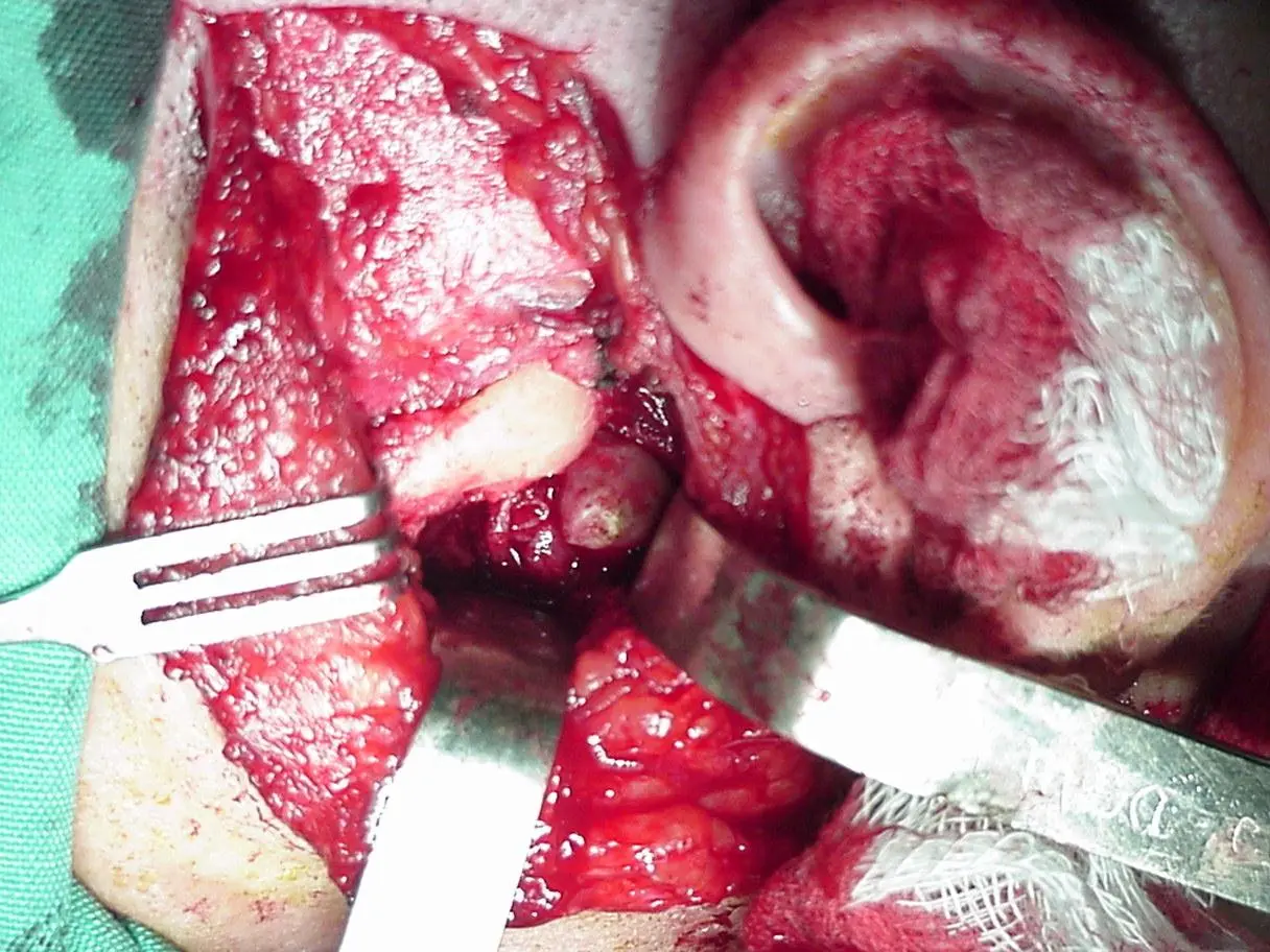
Direct visualization confirmed marked irregularity of the condylar surfaces.
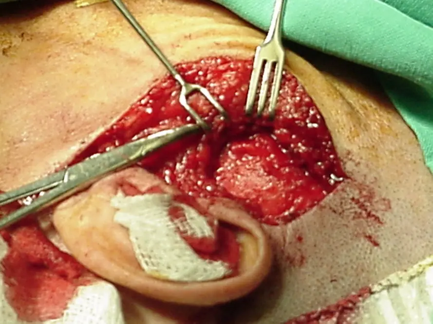
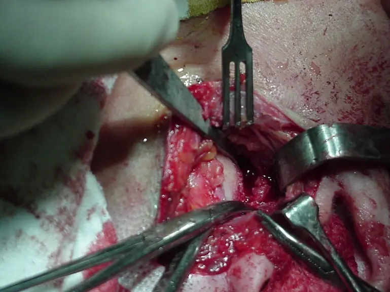
Bilateral preauricular access exposed the temporomandibular joints. The four photographs in this group record the progression from access to direct inspection of the altered condylar anatomy. The operative narrative reports no intraoperative complication.
04 / SELECTIVE CONDYLAR RECONTOURING
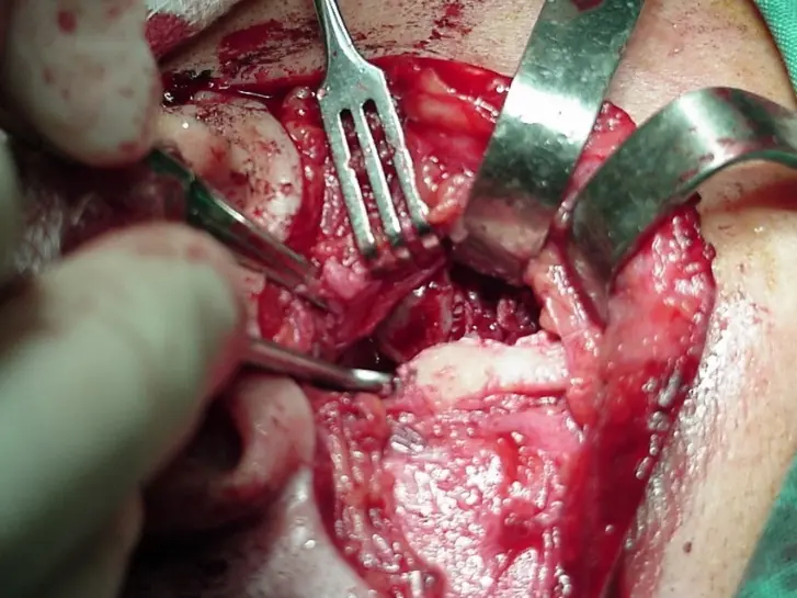
Diamond burs of different shapes were selected according to the available angle of access.
The team performed bilateral condylar arthroplasty using diamond burs of different dimensions and profiles. Instrument selection was adapted to the restricted preauricular working angles and the position of each exostotic projection. The intended endpoint was removal of obstructing irregularities and restoration of a more functional condylar contour while retaining the native joint.
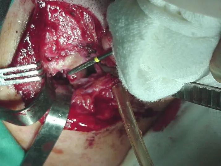
The operation treated bilateral mechanical osseous restriction by targeted reshaping rather than condylectomy or total joint reconstruction. Its value as an archival case lies in this anatomy-preserving operative reasoning. It does not establish comparative superiority, because the record contains no control group or standardized longitudinal outcome measures.
05 / SEVEN-DAY APPEARANCE
Two closure techniques were compared across the bilateral approaches.
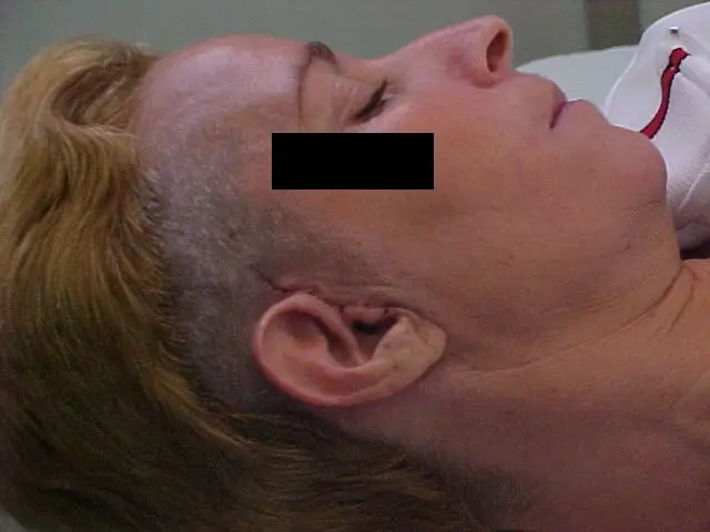
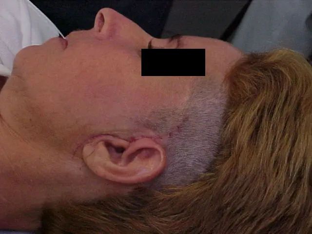
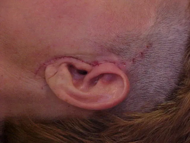
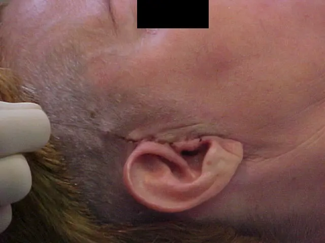
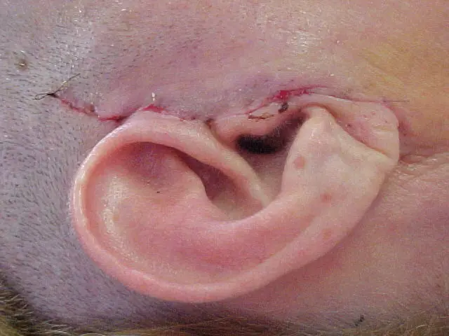
The source identifies this group as the seven-day postoperative visit and suture removal. Different residents performed an intradermal closure on one side and a simple interrupted closure on the other. The team recorded visually similar early aesthetic results and attributed this to the relatively low muscular tension in the preauricular region. That explanation is retained as the team's observation, not as a controlled comparison.
06 / ORIGINAL-SEQUENCE RETURN TO PLANNING
The archive preserves the source order, which next returns to incision marking and closure details.
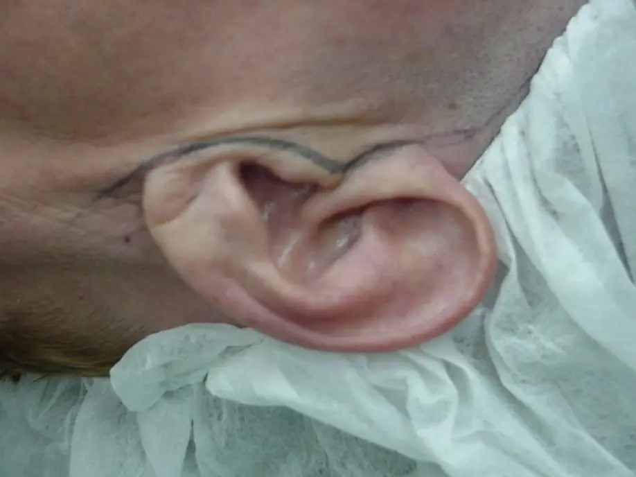
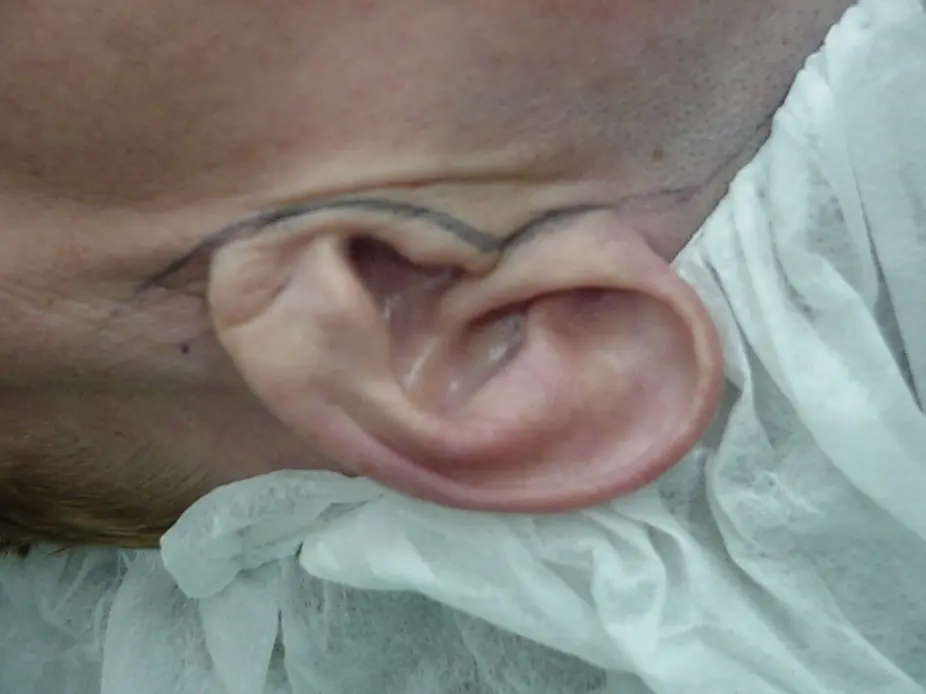
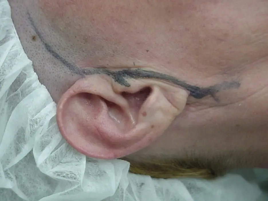
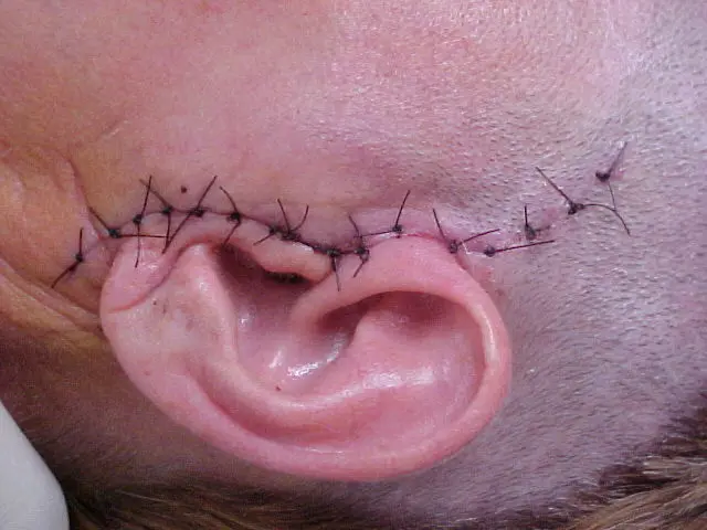
These four photographs appeared after the seven-day group in the original document and remain in that exact position here. They record bilateral preauricular marking with methylene blue, preparation of the operative field, and the two closure patterns later compared by the team.
07 / 24-HOUR RECORD
No facial paralysis was identified in the early postoperative examination.
The final source photograph is identified as 24 hours after surgery. The record states that there was no facial paralysis and that the patient reported disappearance of the preoperative symptoms within the first postoperative day. Because no formal facial-nerve grading, audiological assessment, opening measurement or validated pain score survives, the archive reports this as the treating team's clinical observation.
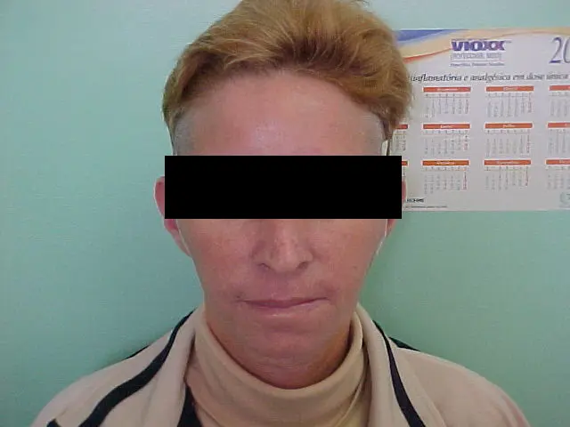
The source further states that the operation proceeded without short- or long-term complications, but it does not identify the duration or schedule of long-term follow-up. The robust conclusion is therefore limited to the documented early clinical recovery and the team's later report of an uncomplicated course.
08 / CONTEMPORARY REFLECTION
The historical procedure sits within a treatment landscape that has since become more diagnostic and less invasive.
Current guidance emphasizes a comprehensive clinical assessment, appropriate imaging and initial nonsurgical care for temporomandibular joint pain and dysfunction. Minimally invasive procedures are generally considered before open arthroplasty, while open surgery is reserved for carefully selected structural disease that remains symptomatic after less invasive management.
Condylar osteophytes and remodeling are imaging signs, not self-sufficient explanations for pain. Their relevance depends on concordance with symptoms, examination and joint mechanics. In this 2002 case, the team linked a prolonged refractory clinical syndrome with bilateral obstructive osseous morphology and chose targeted reshaping of the native condyles. The immediate response is notable, but the missing quantitative and long-term data prevent causal or generalizable claims.
This case demonstrates a disciplined surgical idea: when a discrete anatomical obstruction is judged responsible for mechanical dysfunction, remove only the obstructing morphology and preserve as much native joint structure as possible. Contemporary use of that principle requires modern imaging, diagnostic criteria, shared decision-making and documented outcome measurement.