01 / THE PATIENT ARRIVES
A painless mandibular expansion involved the right body, angle and ramus.
A 23-year-old woman without reported comorbidities presented in 2001 to the Oral and Maxillofacial Surgery residency service coordinated by Professor Gino Emilio Lasco. Carlos A. Andreucci, collaborating surgeons and residents participated in her initial assessment.
Examination showed marked extraoral and intraoral swelling of the right mandible. The expansion involved the body, angle and ramus and extended across both buccal and lingual cortices. No dental mobility was recorded, and the patient reported no pain. Local biopsy established the diagnosis of central giant cell granuloma.
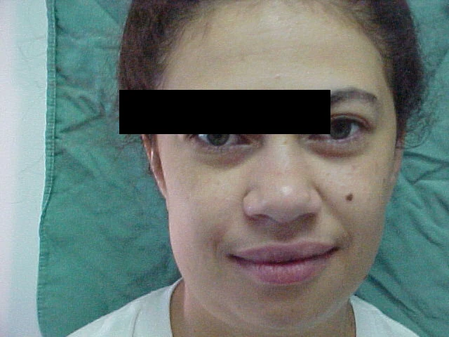
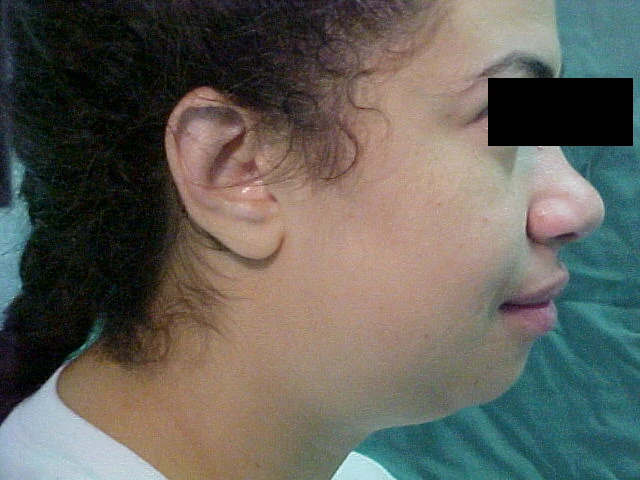
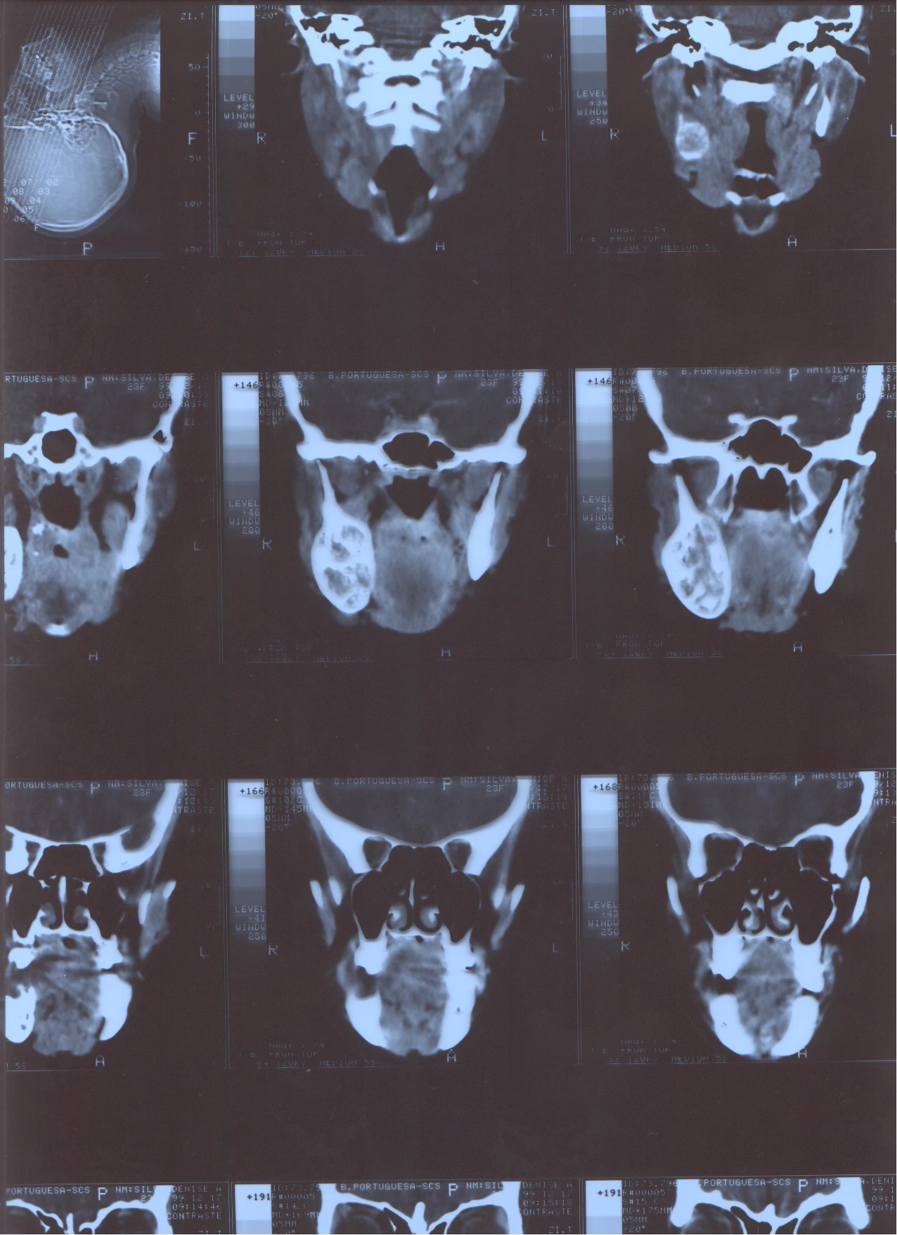
Central giant cell granuloma is a benign intraosseous lesion that can behave locally aggressively. Histology may overlap with giant-cell lesions associated with hyperparathyroidism; contemporary evaluation therefore correlates pathology with imaging and appropriate biochemical testing. Those laboratory data are not present in the surviving record and are not inferred here.
02 / THE DECISION
The reconstruction was designed as a two-stage biological sequence.
Following preoperative examination and general medical assessment, the team planned a partial right mandibulectomy. The lesion's extent made a simple local procedure unlikely to preserve mandibular form. Immediate definitive bone reconstruction was not selected. Instead, the team fabricated temporary metal-acrylic mandibular prostheses in two sizes from direct clinical measurements.
The intention was to remove the lesion while immediately maintaining mandibular length, facial contour and the spatial relationship of the muscles and soft tissues. A second operation would later replace the temporary device with autogenous rib bone after the recipient tissues had matured.
03 / FIRST OPERATION
Occlusion was secured before exposure and partial mandibulectomy.
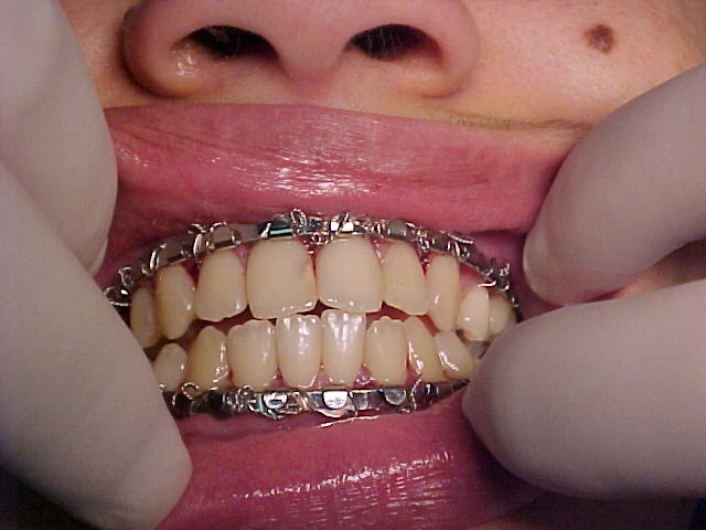
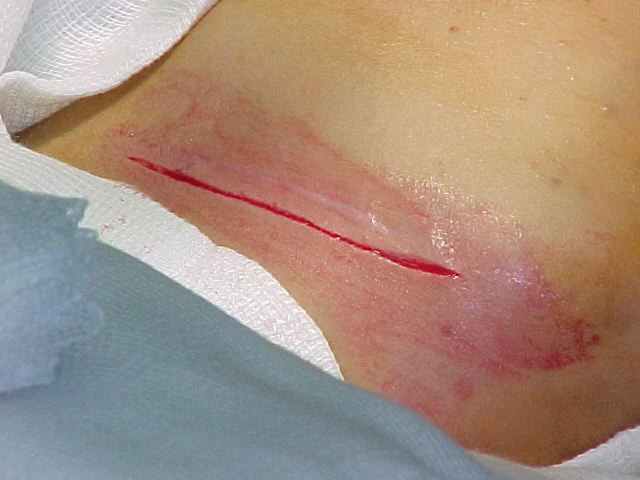
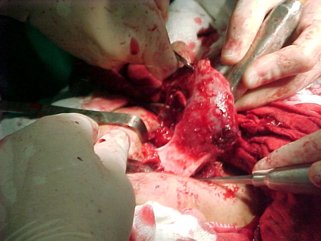
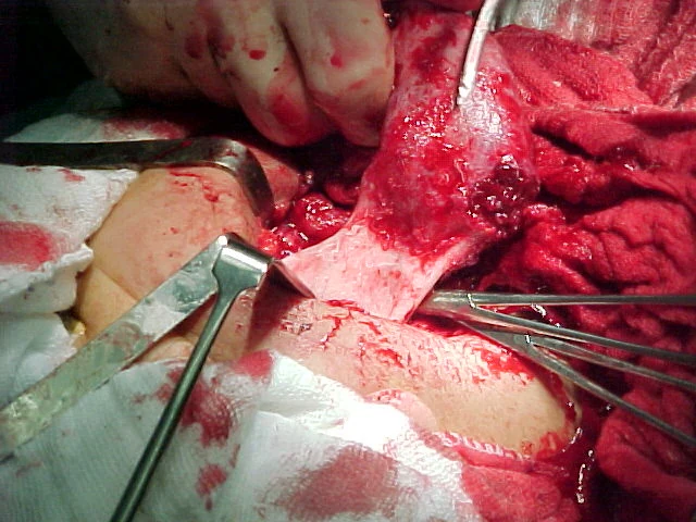
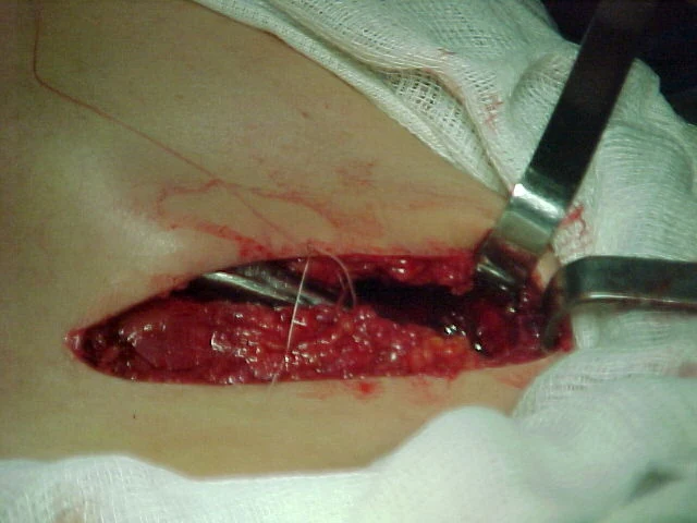
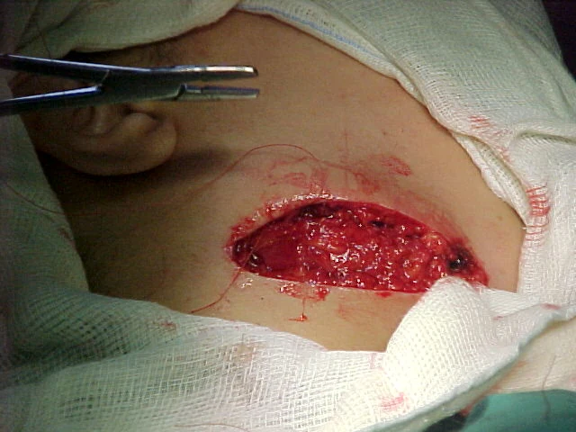
A wide right submandibular approach was marked across the body-angle region. Layered dissection exposed the lesion and adjacent healthy bone. Erich arch bars were applied with 0.0 stainless-steel wire, and maxillomandibular fixation established the occlusal reference before resection.
The team carefully disarticulated the temporomandibular joint while preserving the surrounding tissues, then completed the planned partial mandibulectomy. The operative record emphasizes protection of the articular space and soft-tissue attachments for the temporary reconstruction.
04 / PATHOLOGICAL SPECIMEN
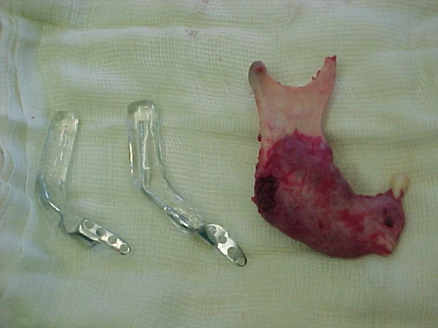
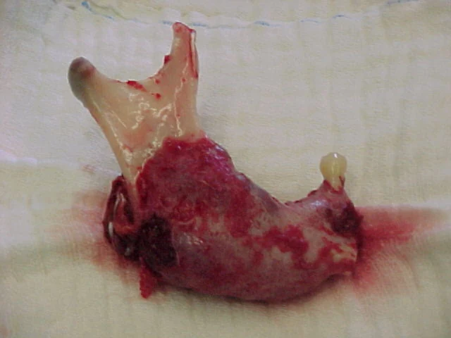
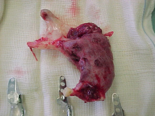
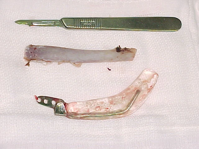
The resected segment preserved the macroscopic record of disease extent.
Two complementary photographs document the resected mandibular segment. The archive retains the biopsy diagnosis of central giant cell granuloma but does not add a histological subtype, margin status or microscopic description that is absent from the source.
05 / TEMPORARY RECONSTRUCTION
The closest prefabricated prosthesis was adapted directly to the defect.
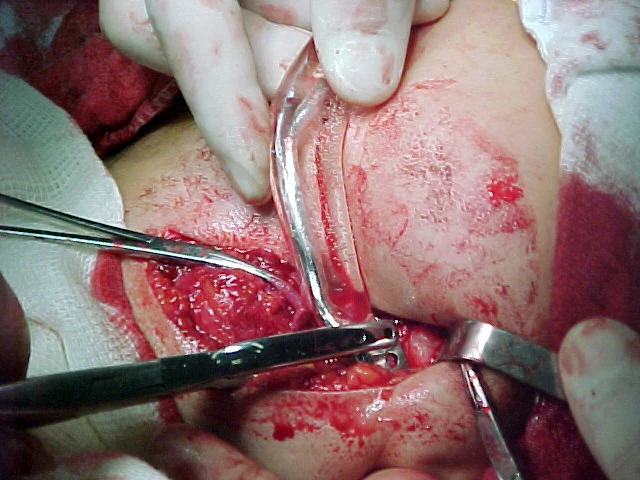
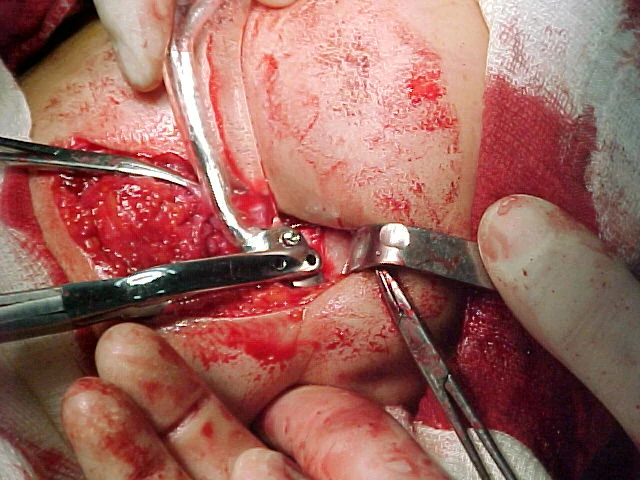
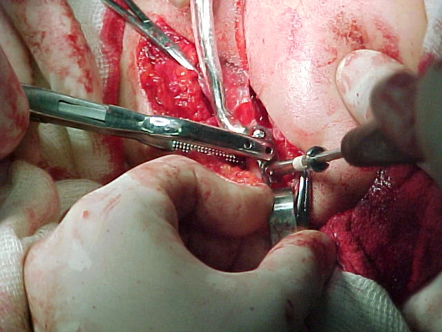
After resection, the team compared the two preoperative prosthesis sizes with the actual defect. The selected device was adapted to restore mandibular continuity and fixed to the remaining anterior mandible with three 10-mm bicortical screws.
The temporary reconstruction was intended to maintain facial form and function while limiting inward displacement and scar contraction of the muscular and soft-tissue envelope. It also preserved an anatomical space for the later autogenous graft.
06 / CLOSURE AND PROTECTED HEALING
Layered closure prepared the tissues for a later biological reconstruction.
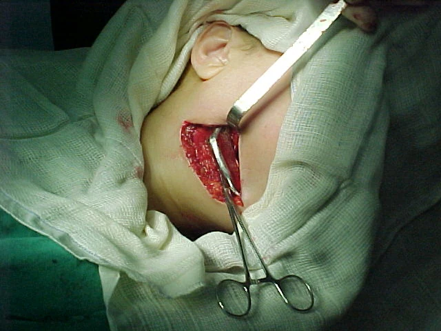
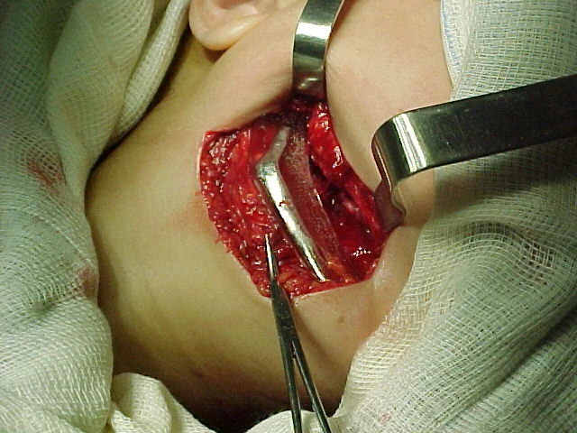
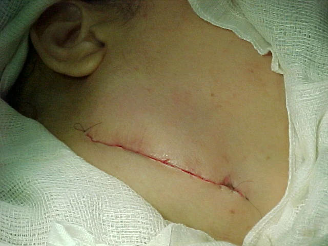
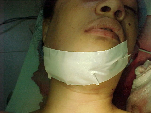
The wound was closed in anatomical layers around the temporary device. The team regarded meticulous coverage and external compression as essential to isolate the future recipient bed, stabilize the restored contour and support uncomplicated healing before the second procedure.
07 / SECOND OPERATION · SIX MONTHS
A free rib graft was harvested for delayed mandibular reconstruction.
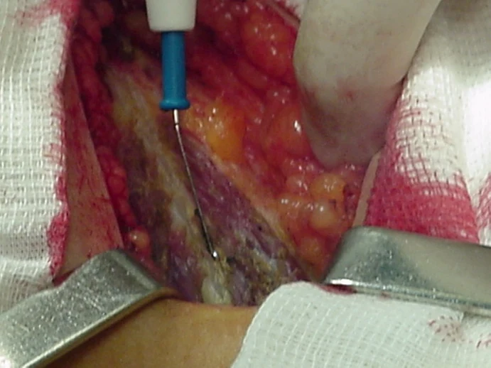
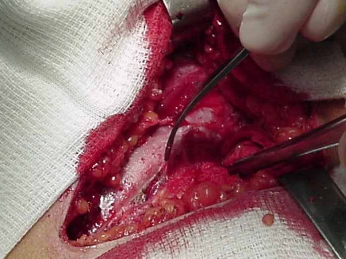
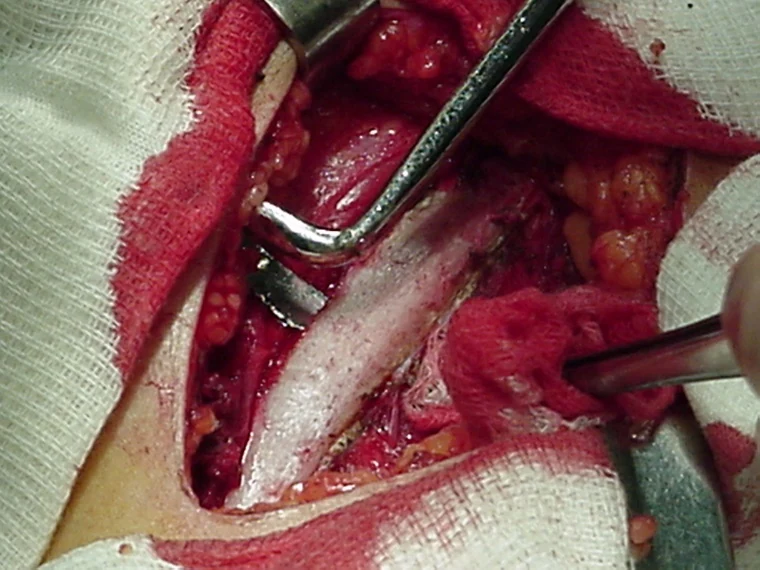
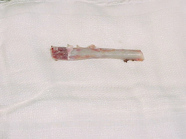
Six months later, a second operation was undertaken. A right floating rib was exposed and removed as a nonvascularized autogenous graft. The graft provided a curved corticocancellous segment that could be shaped to the maintained mandibular space.
08 / THE TEAM'S “PERI-PROSTHESIS” OBSERVATION
The temporary device was removed while its surrounding capsule was preserved.
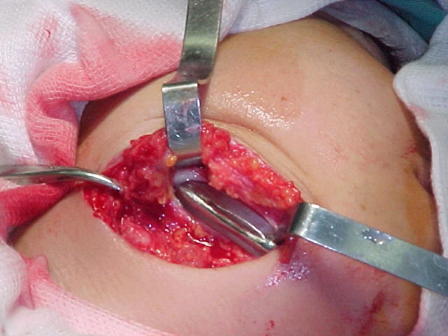
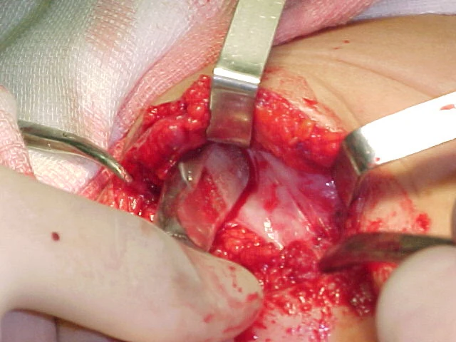
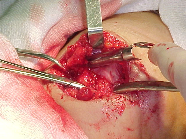
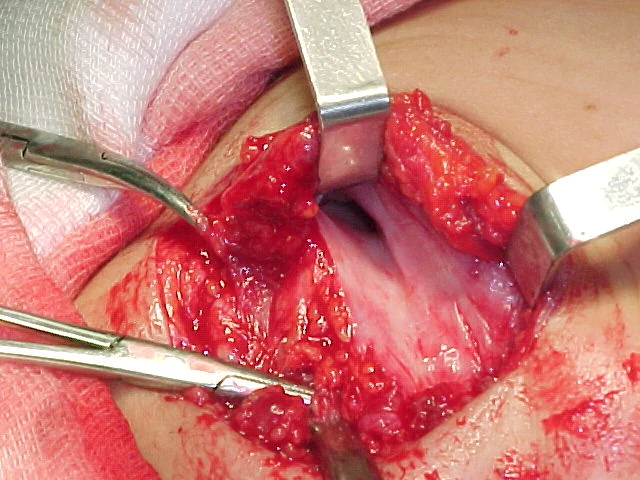
At re-entry, the temporary prosthesis was carefully exposed and removed. The team deliberately preserved the fibrous capsule that had formed around it. Carlos A. Andreucci later used the descriptive term peri-prosthesis for this tissue because its operative appearance and handling recalled a periosteal envelope.
This is historical team terminology, not a histologically validated tissue classification. The surviving record contains no vascular or microscopic study of the capsule. Its documented surgical role was to preserve the reconstructed space and provide a protected envelope around the incoming graft.
09 / BONE-GRAFT ADAPTATION
Bleeding recipient bone and close graft contact guided fixation.
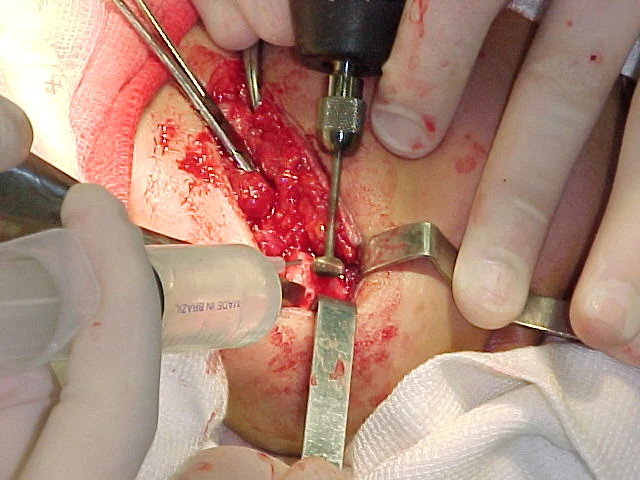
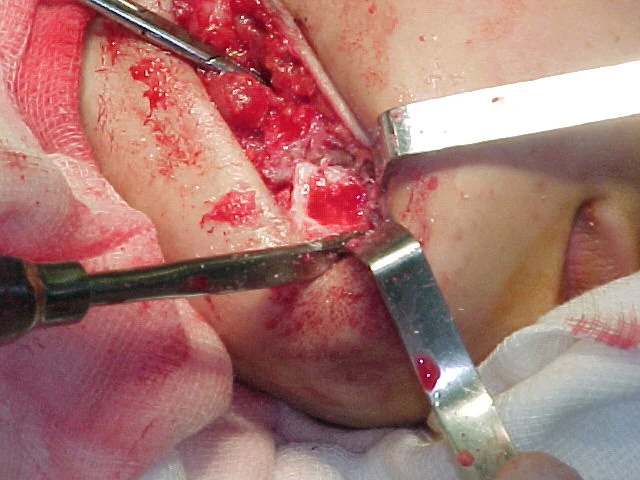
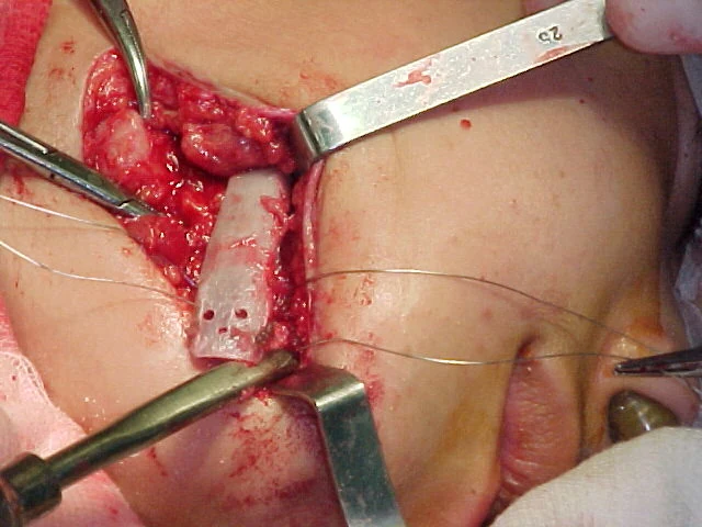
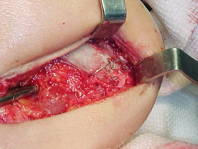
The residual mandibular bone was prepared to expose trabecular bleeding surfaces. The rib graft was contoured for close contact with the recipient mandible and placed inside the preserved soft-tissue envelope. Surgical steel wire fixation was selected to stabilize the graft while reducing the likelihood that later screw removal would be required.
The team believed that maintaining a vascularized soft-tissue cover around the graft would protect it during incorporation and remodeling. This represents the operative rationale recorded by the team; the archive does not claim that graft revascularization or remodeling was independently measured.
10 / POSTOPERATIVE COURSE AND OUTCOME
No complication was recorded, and the case remained in follow-up for five years.
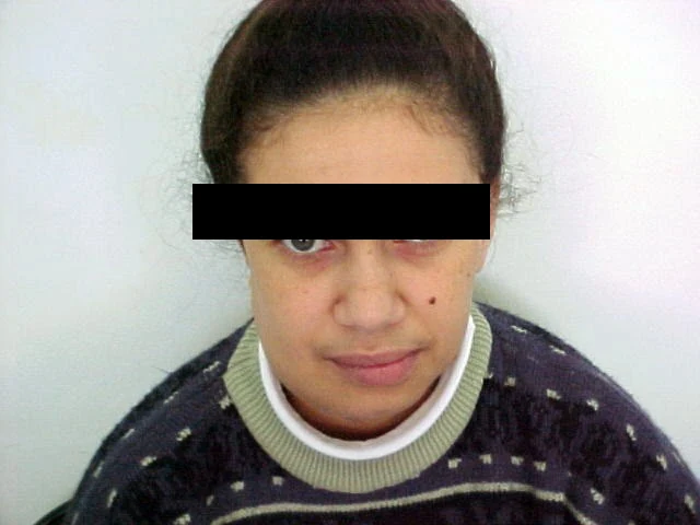
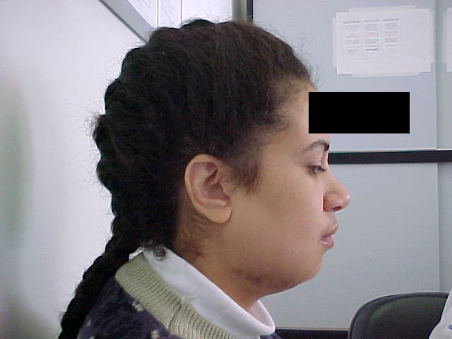
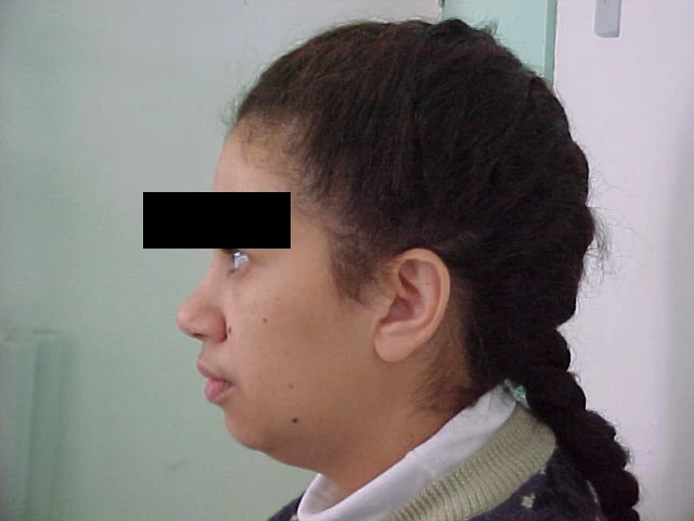
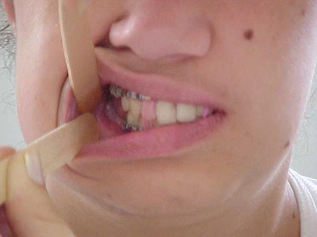
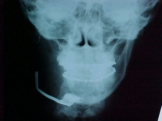
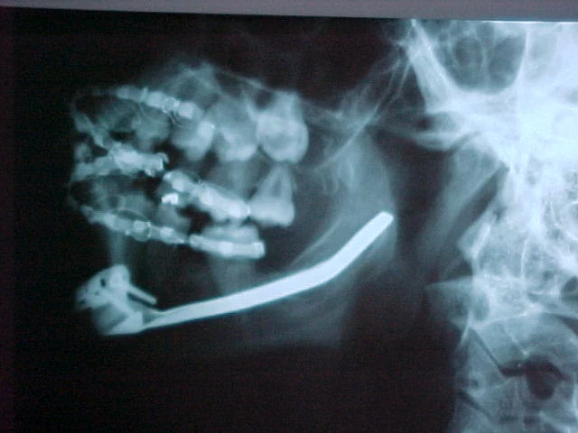
The source reports no complication after either operation. The patient maintained function, facial continuity and quality of life during the five-year recorded follow-up. The first four images below document the early postoperative appearance identified in the original document as 15 days after the first operation. The final two radiographs belong to the supplied outcome sequence, but their exact dates are not identified in the surviving text.
11 / WHY THIS CASE IS DISTINCTIVE
The temporary prosthesis was used as a spatial and soft-tissue strategy, not as the endpoint.
The innovation was the planned sequence. The first-stage prosthesis immediately restored length and contour after a major resection. Six months of healing allowed the soft tissues to organize around a stable geometry. At the second operation, the device was exchanged for autogenous bone without surrendering the space created during the first procedure.
Contemporary reconstruction may consider vascularized bone transfer, patient-specific plates, virtual planning and, in selected central giant cell granulomas, intralesional or systemic therapies. Published reviews confirm that treatment remains heterogeneous and depends on lesion behavior, extent, morbidity and recurrence risk. Those modern options provide context, but they do not rewrite the 2001 decision.
The preserved capsule resembles principles later emphasized in staged reconstructive techniques, but this archive does not label it an induced membrane or claim biological equivalence. No membrane histology, perfusion data or controlled comparison survives for this case.