01 / INTRODUCTION AND HISTORICAL FOUNDATION
Sixty years of technical evolution led to a method centered on anatomy, nerve protection and functional stability.
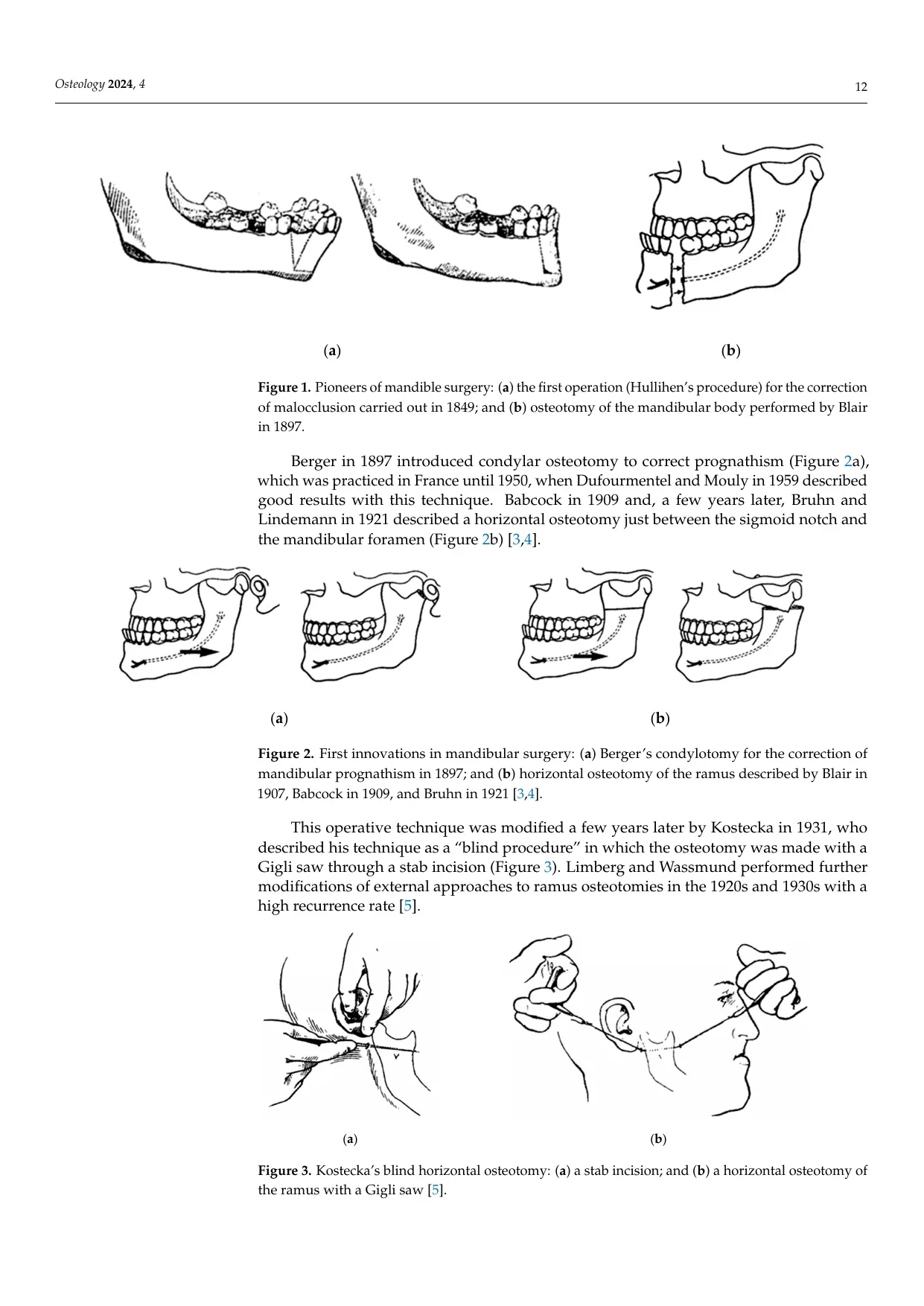
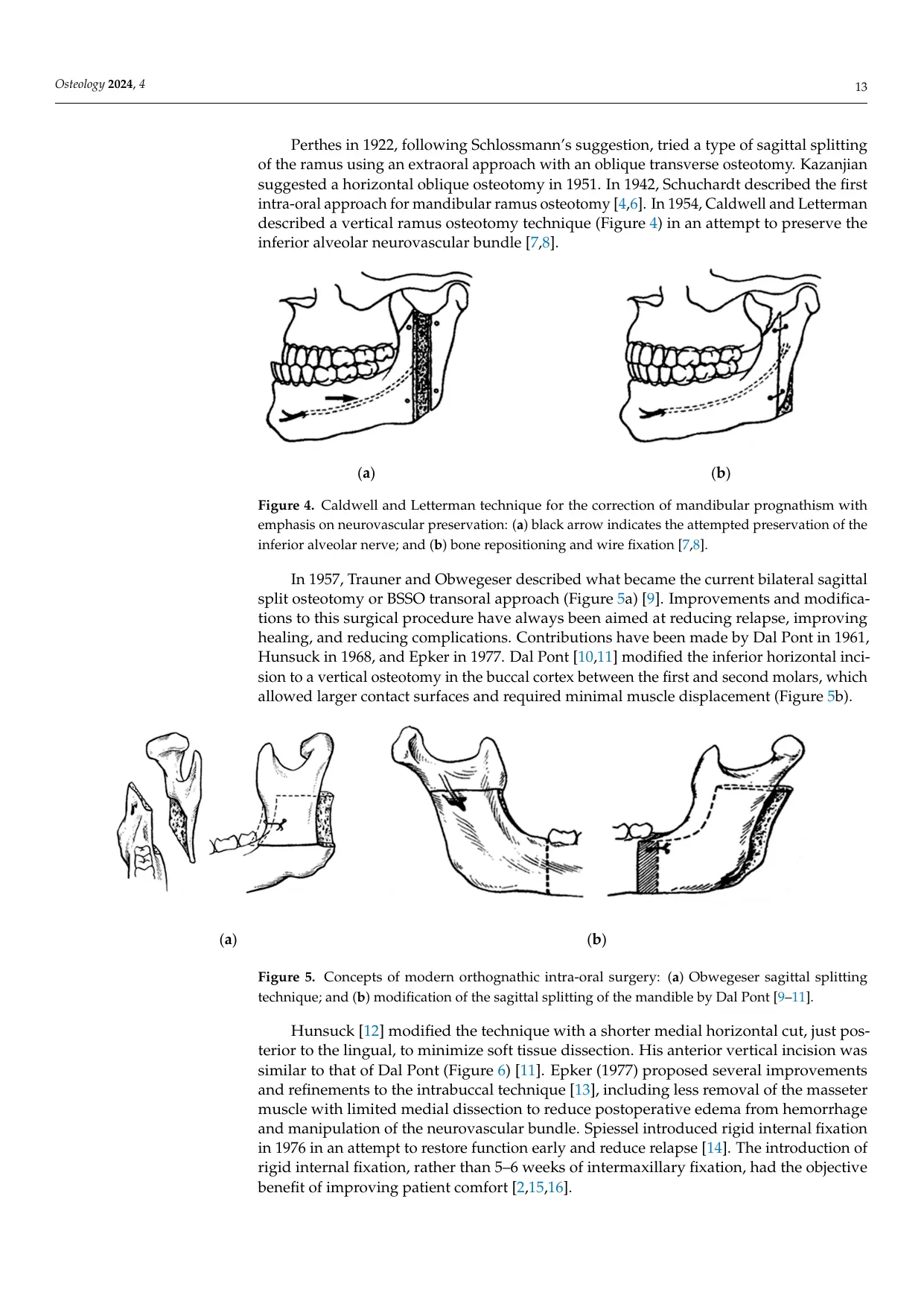
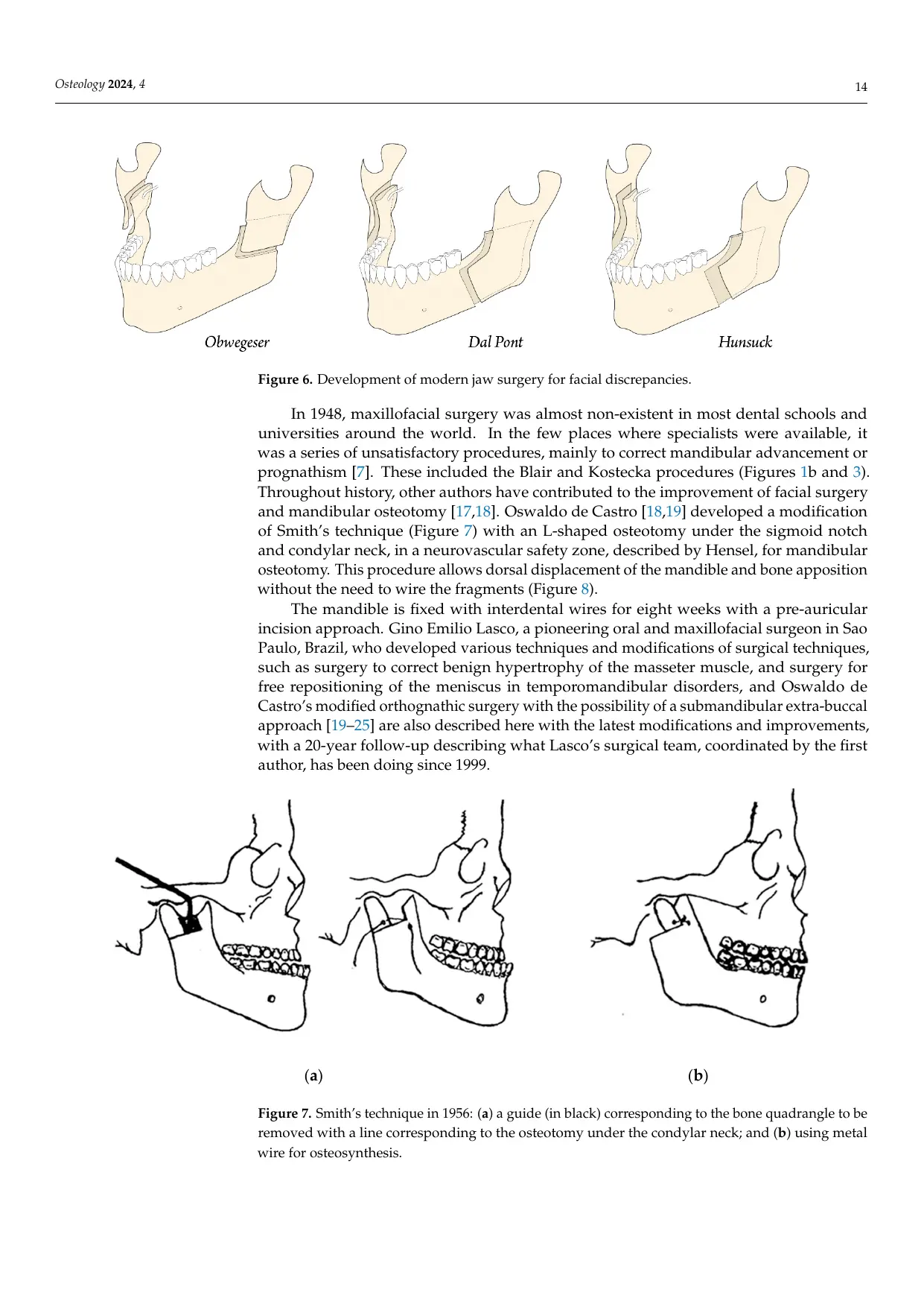
Mandibular orthognathic surgery evolved through body osteotomies, condylotomies, horizontal and vertical ramus procedures, sagittal split techniques and many later modifications. The historical review shows that each innovation attempted to reduce relapse, protect the inferior alveolar neurovascular bundle, improve healing and obtain a stable occlusion with an acceptable facial result.
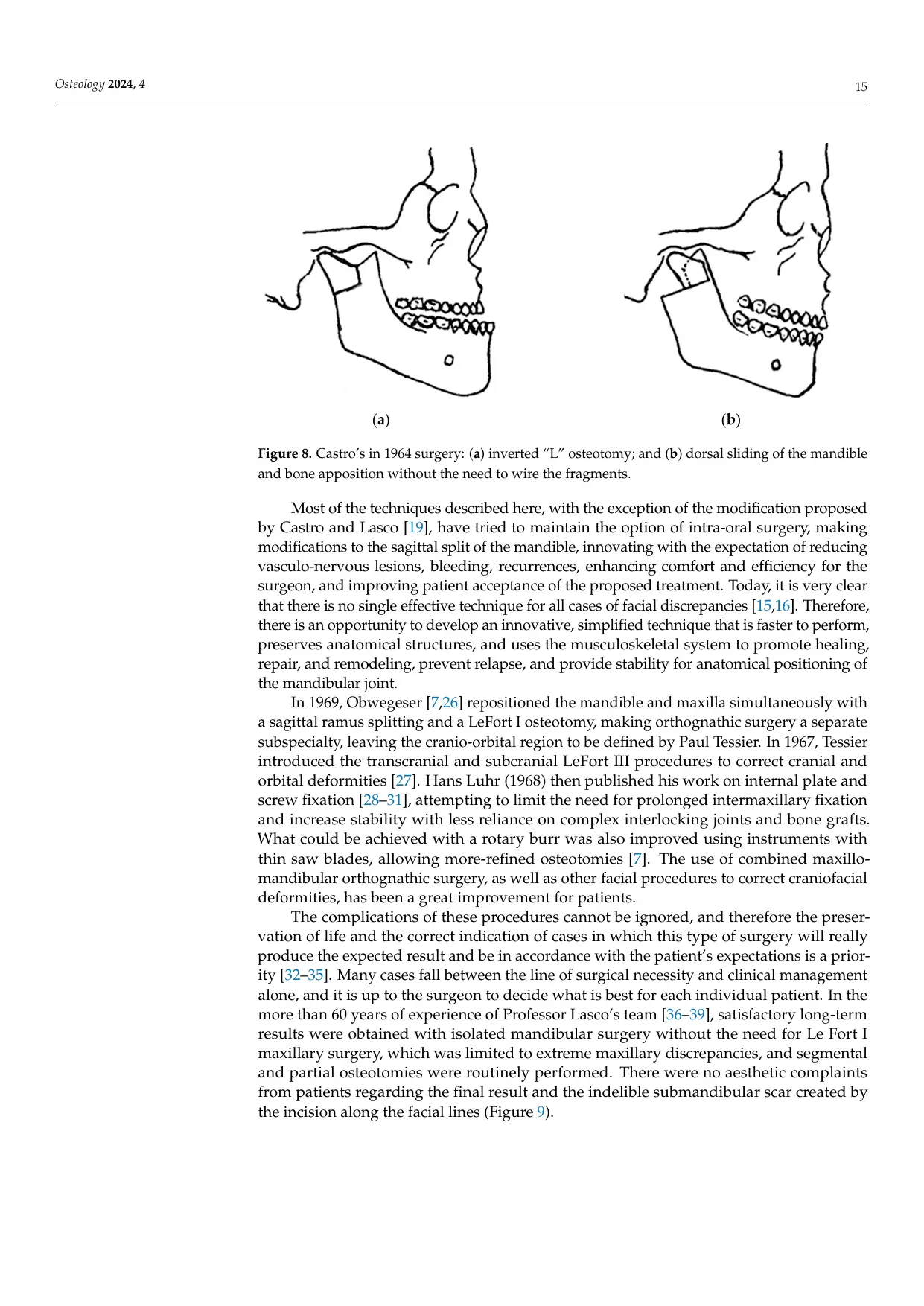
Professor Lasco's school retained the useful biomechanical principles of earlier ramus osteotomies while modifying the access, bone cut, fragment control and postoperative positioning. The objective was not simply to move the mandible. It was to reposition the mandible while preserving vascular and neural structures, maintaining the condyle in a functional relationship with the glenoid fossa, and allowing bone repair without routine rigid internal fixation.
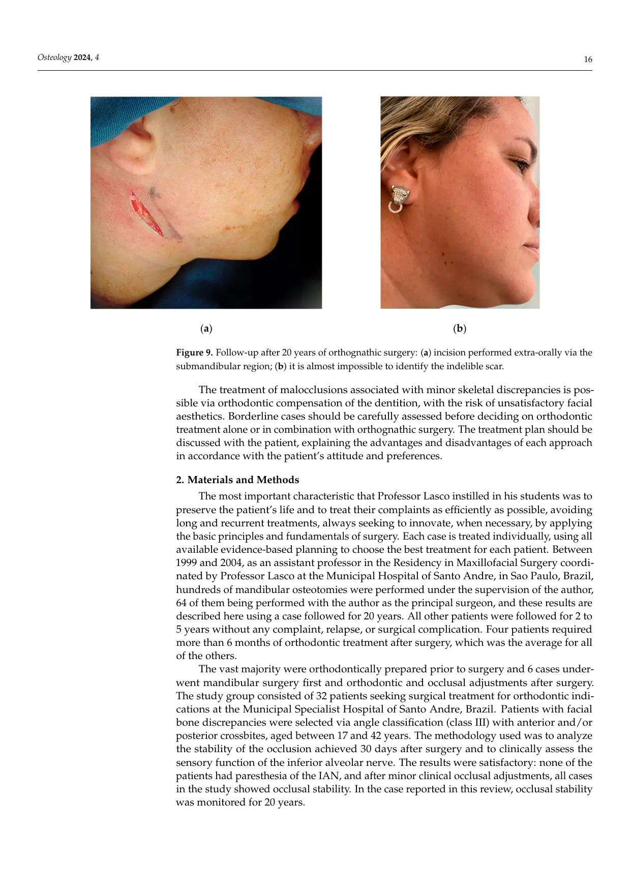
The images below reproduce the original article's review of Figures 1 to 9. They place Professor Lasco's technique within the lineage from the first mandibular osteotomies to modern orthognathic surgery.
02 / MATERIALS, CASE AND SURGICAL TECHNIQUE
The operation combined model planning, a limited submandibular approach, a modified inverted L osteotomy and controlled functional repositioning.
Between 1999 and 2004, hundreds of mandibular osteotomies were performed within the residency coordinated by Professor Lasco. The reported study group included 32 patients, 17 to 42 years old, with Class III facial bone discrepancies and anterior or posterior crossbites. Sixty-four osteotomies were performed with Carlos A. Andreucci as principal surgeon. Most patients received presurgical orthodontic preparation, while six underwent a surgery-first sequence followed by orthodontic and occlusal adjustment.
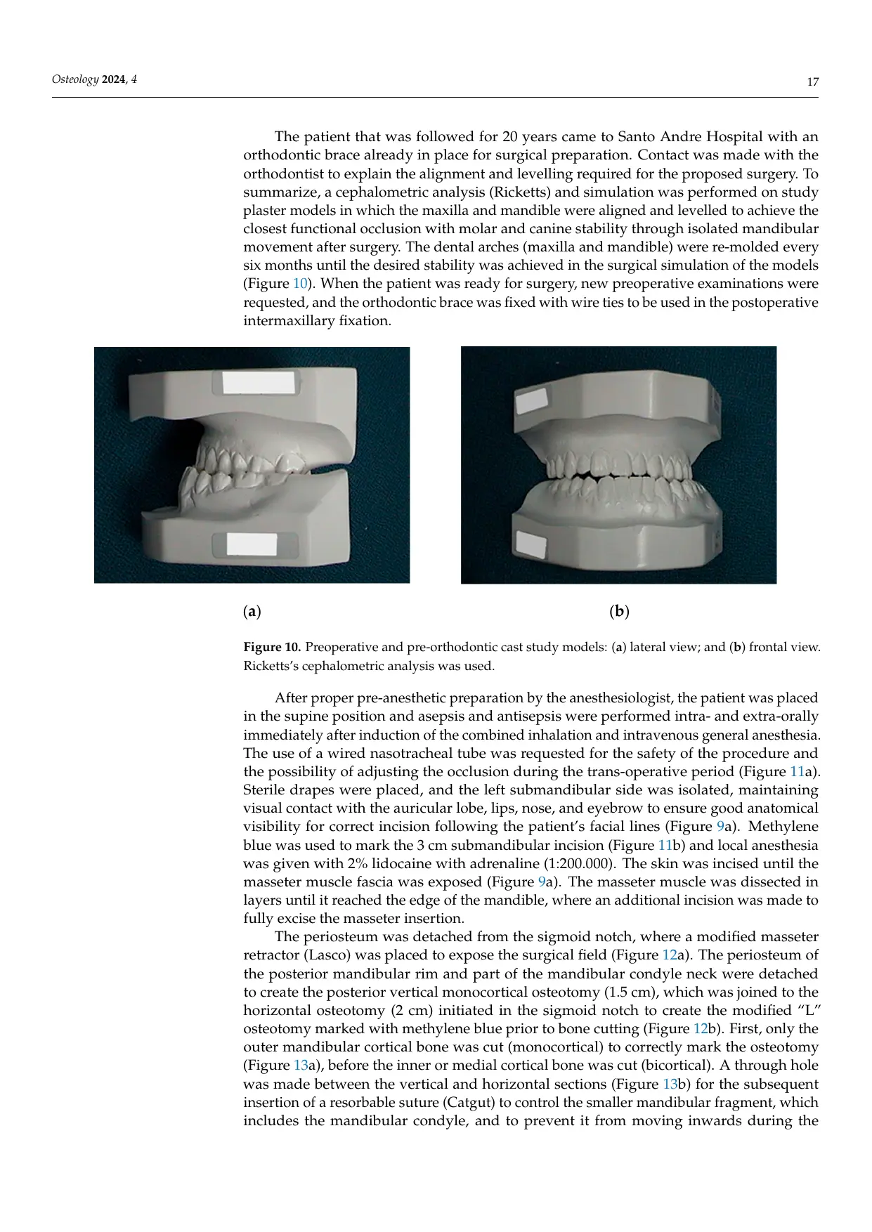
The 20-year case arrived with orthodontic appliances already in place. Ricketts cephalometric analysis and repeated cast-model simulation were used to prepare molar and canine stability through isolated mandibular movement. The maxilla did not require osteotomy, and genioplasty was not added.
Professor Lasco's operative sequence
- Safe access.Nasotracheal general anesthesia permitted intraoperative occlusal assessment. A carefully planned 3 cm submandibular incision followed the facial lines.
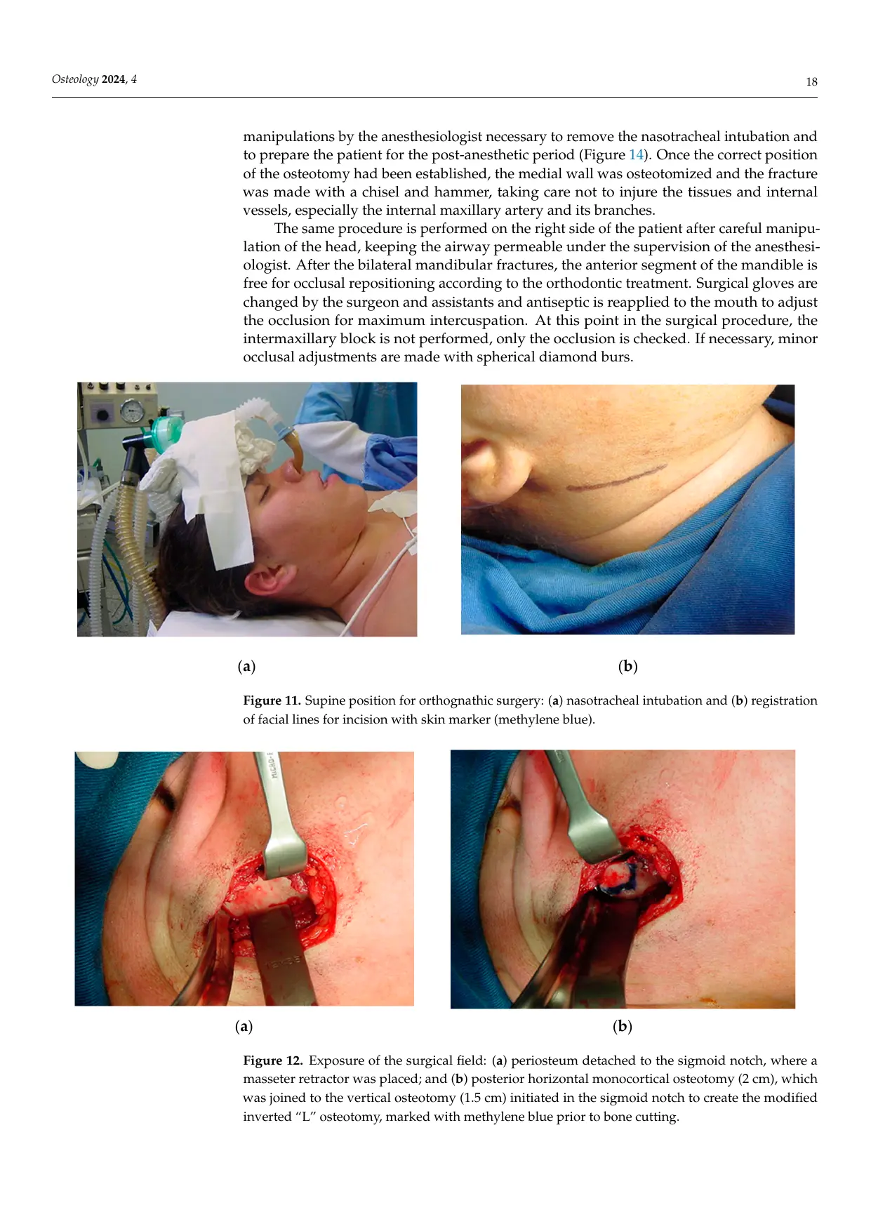
- Muscular and periosteal exposure.The masseter was dissected in layers and released at its insertion. A modified Lasco masseter retractor exposed the sigmoid notch.
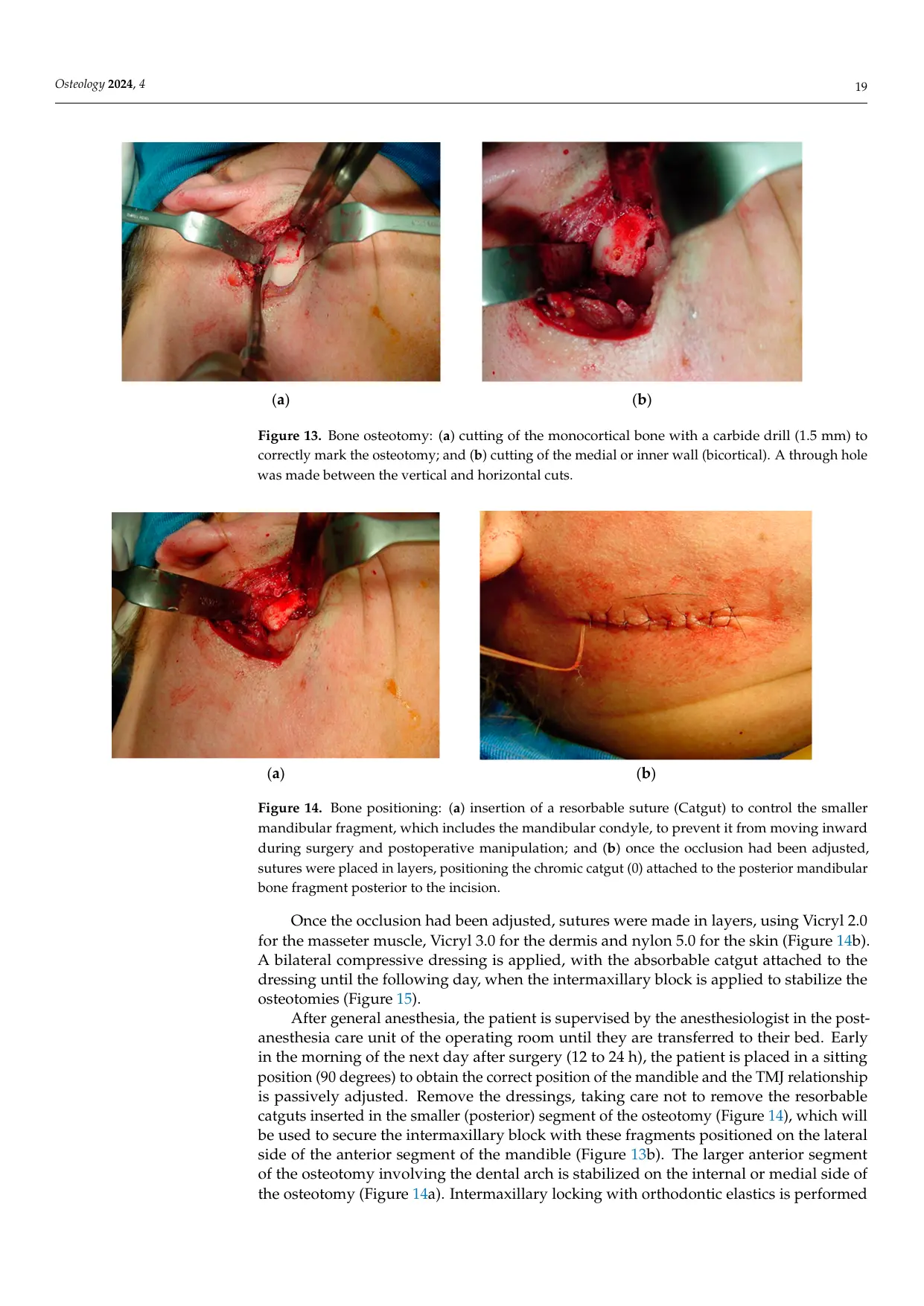
- Modified inverted L osteotomy.A 2 cm horizontal cut beginning at the sigmoid notch joined a 1.5 cm posterior vertical cut. The outer cortex was marked first, followed by bicortical completion under direct protection of the internal tissues and vessels.
- Fragment control.A transosseous hole accepted a resorbable catgut suture. This controlled the smaller condylar fragment and reduced the risk of medial displacement during surgery and recovery.
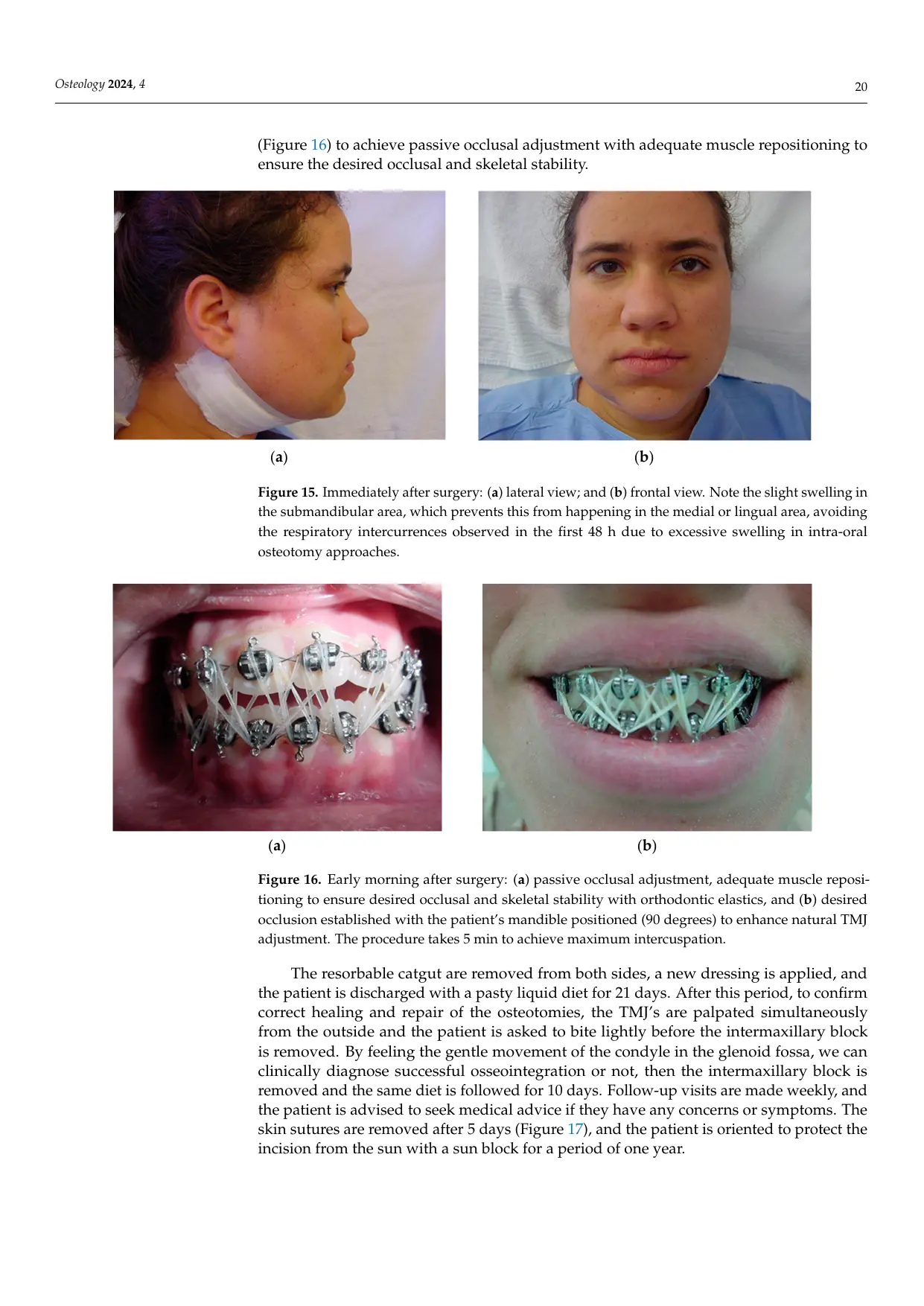
- Occlusal and functional positioning.After bilateral mobilization, the anterior mandibular segment was repositioned to the planned occlusion. The next morning, with the patient seated at 90 degrees, the condyles, muscles and occlusion were passively adjusted before elastic maxillomandibular stabilization.
- Healing without rigid fixation.The intermaxillary block was maintained for 21 days, followed by clinical assessment of condylar movement, progressive diet and orthodontic finishing.
03 / RESULTS
Stable occlusion, preserved sensation and complete long-term remodeling supported the technique.
All patients in the study group obtained occlusal stability after minor postoperative adjustments. No patient in the group developed inferior alveolar nerve paresthesia. Across the 64 osteotomies reported by the author, there were no recurrences, no malocclusions that could not be corrected by occlusal adjustment, no significant neurovascular injury and no significant scarring.
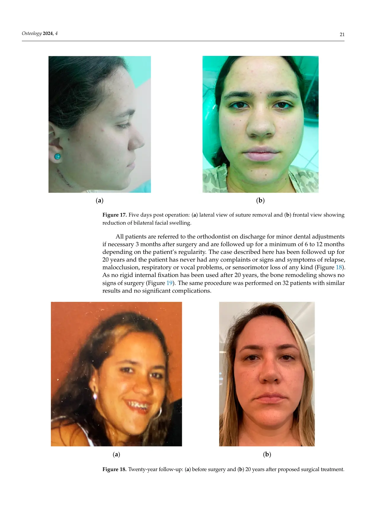
In the classical case, the planned occlusion remained stable throughout more than 20 years of follow-up. The patient reported no relapse, respiratory or phonetic problem, or sensorimotor loss. Preoperative temporomandibular pain, clicking and weekly headaches were no longer reported after functional correction. The long-term panoramic radiograph showed remodeling without visible evidence of fixation hardware because rigid internal fixation had not been used.
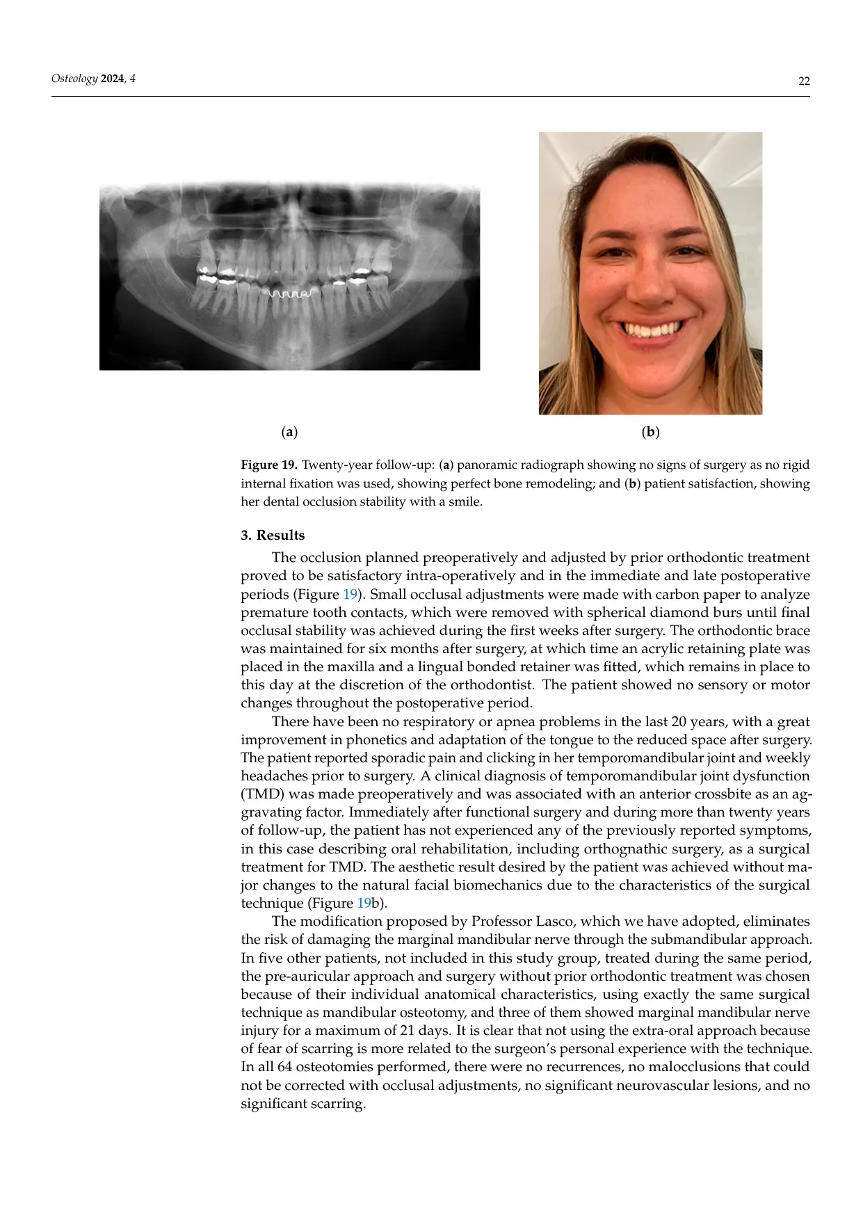
The continuity of the work lies in combining surgical access, osteotomy geometry, fragment control, occlusion, muscular balance and condylar position as one biomechanical treatment. Hardware was not treated as the objective. Stable function and biological repair were the objectives.
04 / PUBLISHED SOURCE
Read the complete peer-reviewed article.
This classical case edition adapts Sections 1, 2 and 3 of the open-access article and preserves all Figures 1 to 19 within the original page plates. The complete publication includes the later discussion, conclusions and references.
Osteology · 2024 · Open accessAndreucci, C.A. Sixty Years of Innovation in Biomechanical Orthognathic Surgery: The State of the Art and Future Directions.Open the article home page ↗