01 / THE PATIENT ARRIVES
Five years of painless swelling had become a structural mandibular problem.
The patient was a 35-year-old man without reported comorbidities who presented in 1975 during the residency program directed by Professor Gino Emilio Lasco. His complaint was painless swelling in the right submandibular region. According to the history obtained from the patient, the swelling had evolved over approximately five years.
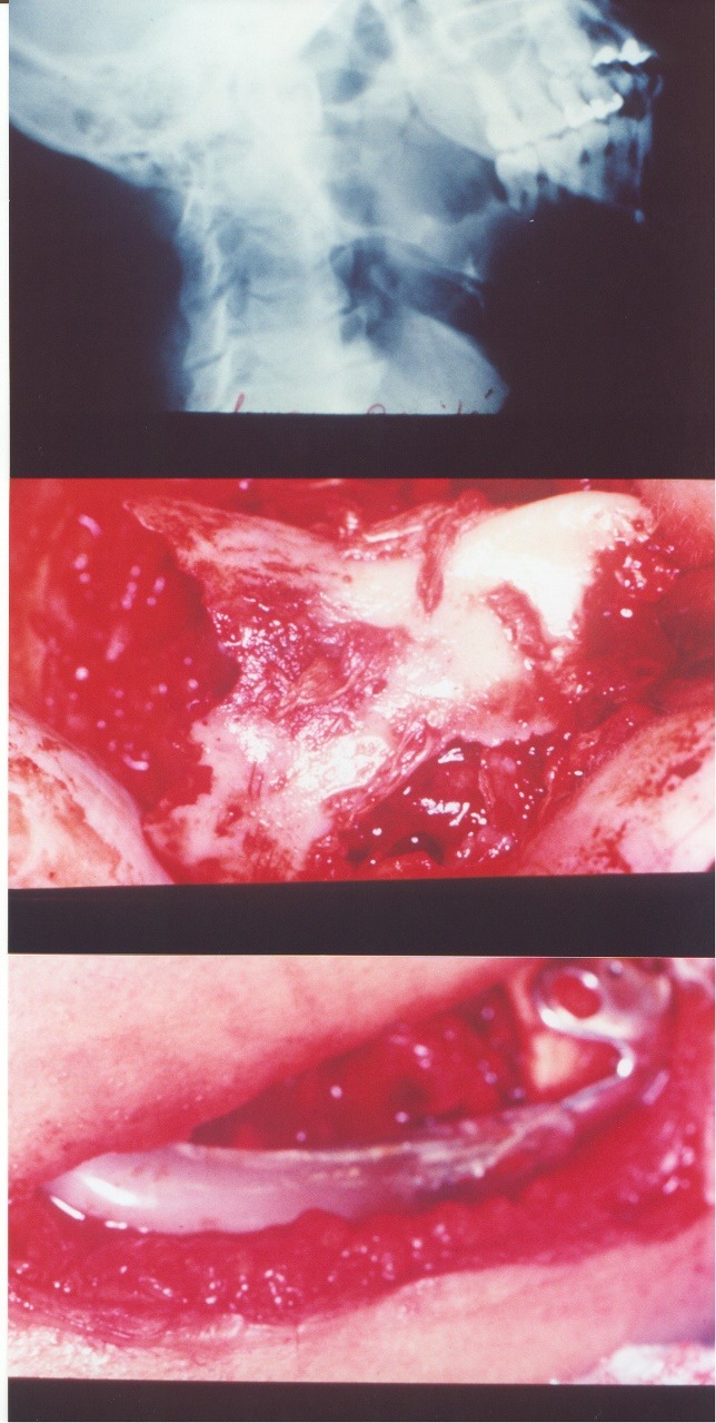
Clinical examination documented indurated lingual and buccal swelling and mobility of teeth 46, 47 and 48. The radiographic examination supported the team's initial working diagnosis of ameloblastoma.
02 / DIAGNOSTIC CONFIRMATION
The clinical and radiographic hypothesis was confirmed as ameloblastoma.
The source identifies ameloblastoma as the initial diagnosis supported by the radiograph. During surgery, tissue was removed for anatomopathological examination before the resection proceeded. The archive curator confirms that the pathology report established the diagnosis of ameloblastoma.
The microscopic description, histological subtype, lesion measurements, margin report and complete radiographic series are not currently available. This public reconstruction therefore records the confirmed diagnosis without inventing a subtype or unsupported pathological detail.
The confirmed diagnosis is included from the curator's record. Pathological details that are not currently available remain explicitly undescribed.
03 / PLANNING BEFORE DIGITAL IMAGING
Three manually fabricated prosthetic sizes prepared the team for uncertainty.
The surgical plan included laboratory fabrication of three differently sized prosthetic models using chromium-molybdenum and methyl methacrylate materials. Direct extraoral and intraoral facial measurements were used because contemporary three-dimensional imaging, virtual planning and patient-specific manufacturing were unavailable.
The three sizes provided a practical safety range. After the lesion was exposed and the partial mandibulectomy completed, the models could be compared directly with the defect before the definitive temporary prosthesis was selected.
04 / OPERATIVE RECORD
Resection and immediate maintenance of mandibular continuity.
The operation used a mandibular incision extending through the angle and mental regions. The tissue planes were dissected to expose the full lesion. A specimen was removed for anatomopathological examination, followed by partial mandibulectomy.
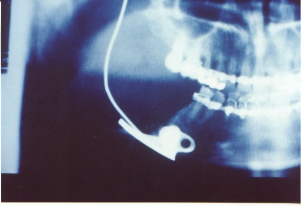
The available prosthetic sizes were compared with the resulting defect. The selected prosthesis was fixed with two bicortical screws, each reported as 10 mm long and 2.0 mm in diameter. Closure was completed in layers.
05 / THE RESECTED SPECIMEN
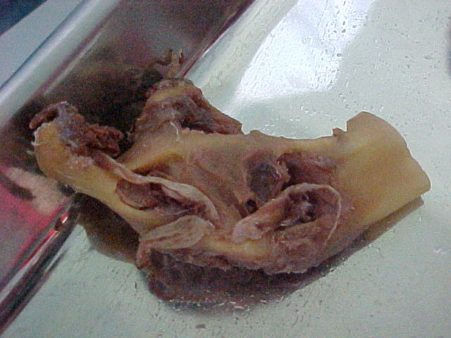
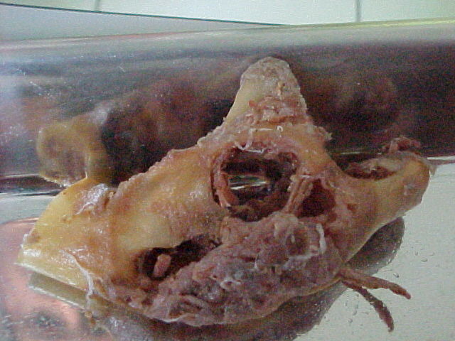
The macroscopic record preserved the extent of the mandibular lesion.
The document includes two later macroscopic views of the resected mandibular specimen. They are retained together because they show complementary surfaces of the same historical pathological record.
06 / STAGED RECONSTRUCTION AND OUTCOME
The temporary prosthesis preserved the region until biological reconstruction.
The historical narrative reports five years of follow-up without recurrence. Oral rehabilitation during the documented course used removable partial prostheses.
One year after the initial operation, the temporary prosthesis was removed and the mandibular region was reconstructed with a rib graft. The record and images of that second operation are not currently available. The transplant is therefore documented as a confirmed later stage, but its technique and result are not reconstructed or illustrated on this page.
The initial operation controlled the lesion and maintained mandibular form with an immediately available temporary reconstruction. Definitive biological reconstruction was deliberately staged for a later operation.
07 / HISTORICAL REFLECTION
Adaptability was engineered into the plan before the incision.
The central lesson is not only the prosthesis itself. The team anticipated uncertainty by fabricating three sizes, then selected the closest reconstruction after direct visualization of the true defect. In a period without CT-based planning or patient-specific implants, redundancy became a practical planning tool.
Today, the lesion would ordinarily be characterized with cross-sectional imaging and histopathological diagnosis, while virtual surgical planning, cutting guides, patient-specific reconstruction and contemporary microvascular or nonvascular graft options could be considered according to the defect and patient. That modern reflection does not replace the historical rationale or imply that one present-day pathway is appropriate for every case.