01 / THE PATIENT ARRIVES
A severe skeletal discrepancy with functional complaints.
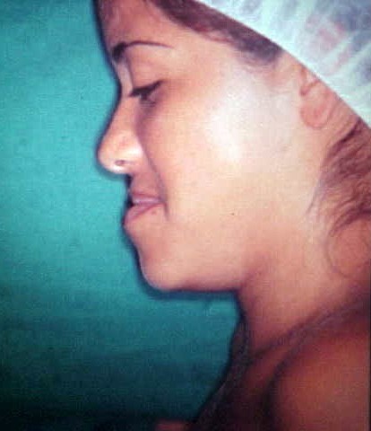
The patient was an 18-year-old woman with severe mandibular prognathism. The supplied clinical record documents atypical swallowing, headaches and difficulty with speech production.
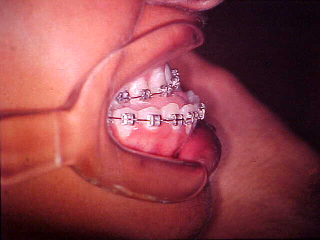
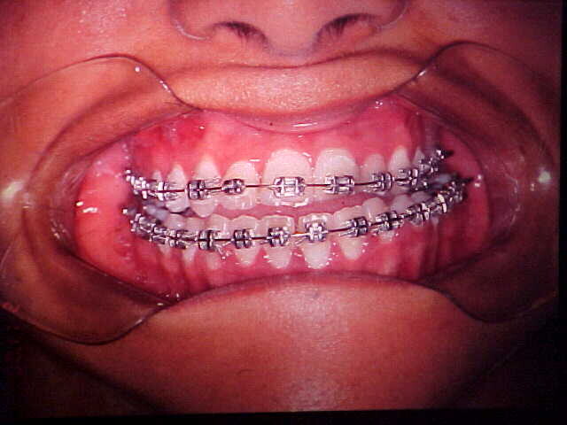
Presurgical orthodontic treatment was indicated to prepare the dental arches for surgical correction. The surviving record does not provide a complete cephalometric analysis or a formal differential diagnosis, so this page preserves the diagnosis and treatment sequence exactly at the level supported by the source.
02 / MODEL SURGERY AND FUNCTIONAL PLANNING
Test the movement and occlusion before entering the operating room.
Plaster casts were mounted on an articulator. The team performed the planned skeletal movement on the models to verify that the postoperative segments could produce a stable occlusal relationship and a functionally acceptable bite.
This laboratory step allowed the intended mandibular repositioning to be evaluated before surgery and provided an occlusal reference for intraoperative adjustment.
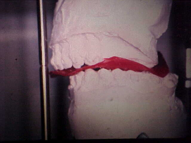
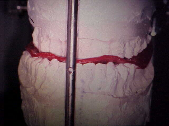
The original document does not include measurements of the planned setback, cephalometric tracings or details of the orthodontic sequence. No values have been reconstructed or inferred for this public edition.
03 / OPERATIVE DECISION
An extraoral concept adapted to an intraoral route.
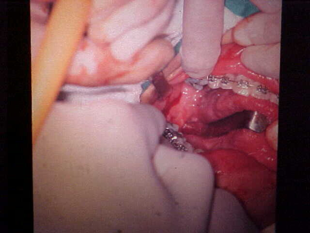
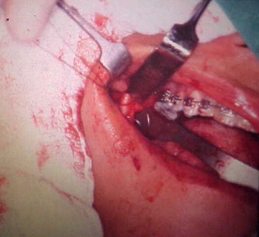
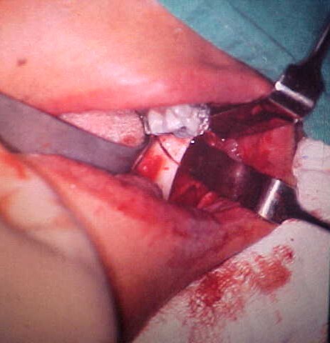
The selected operation was a bilateral horizontal osteotomy of the mandibular rami performed through an intraoral approach. The record describes this as an adaptation of the extraoral Babcock technique, made possible by instruments that allowed more precise cutting and surgical access.
The objective was to reposition the mandible according to the planned occlusion while avoiding an external surgical access. The source records the choice but does not list every alternative considered by the team at that time.
04 / OPERATIVE RECORD
Bilateral horizontal ramus osteotomies with wire stabilization.
The bilateral horizontal osteotomies were completed while respecting the anatomical limits visible through the intraoral access. Rigid internal fixation was not used. A surgical steel wire maintained the osseous segments in position.
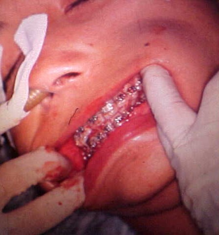
Maxillomandibular fixation and immobilization were obtained through the orthodontic brackets, which had been adapted and reinforced for the postoperative period.
05 / OCCLUSAL ADJUSTMENT AND RECOVERY
Twenty-one days of fixation followed by functional consolidation.
The occlusion was adjusted and tested in the operating room. The source states that definitive immobilization was completed 24 hours later in the outpatient clinic while the patient was seated, allowing the team to reassess condylar position at the temporomandibular joints and muscular adaptation before final fixation.
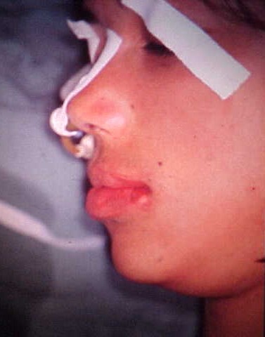
Maxillomandibular fixation was maintained for 21 days. After release, functional osseous consolidation was recorded. Final discharge was documented at three months, after completion and removal of the orthodontic treatment described in the source.
06 / WHAT THE RECORD PRESERVES
Occlusion, joint position and muscle balance were treated as one system.
The case illustrates Professor Lasco's team's emphasis on planning the occlusion before surgery, verifying it again in the operating room and reassessing the patient after the immediate postoperative muscular response. The 24-hour seated adjustment was recorded as part of respecting temporomandibular joint repositioning and muscular readaptation.
From a present-day perspective, three-dimensional imaging, virtual surgical planning, digital splints and contemporary rigid fixation could provide additional planning and stabilization options. This reflection does not replace the original rationale. The historical value of the case lies in how model surgery, intraoral access, limited wire stabilization and controlled immobilization were combined to achieve the reported functional consolidation.